

Tucker Marr’s life changed forever last October.
He was on his way to a wedding reception when he fell down a steep flight of metal stairs, banging the right side of his head so hard he went into a coma.
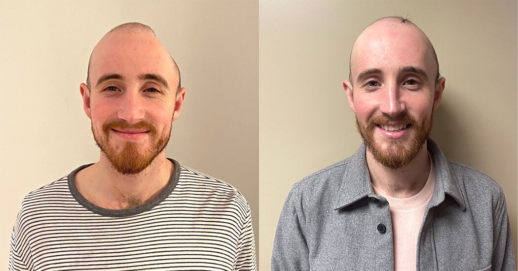
He’d fractured his skull, and a large blood clot formed on the left side of his head. Surgeons had to remove a large chunk of his skull to relieve pressure on his brain and to remove the clot.
“Getting a piece of my skull taken out was crazy to me,” Mr. Marr said. “I almost felt like I’d lost a piece of me.”
But what seemed even crazier to him was the way that piece was restored.
Mr. Marr, a 27-year-old analyst at Deloitte, became part of a new development in neurosurgery. Instead of remaining without a piece of skull or getting the old bone put back, a procedure that is expensive and has a high rate of infection, he got a prosthetic piece of skull made with a 3-D printer. But it is not the typical prosthesis used in such cases. His prosthesis, which is covered by his skin, is embedded with an acrylic window that would let doctors peer into his brain with ultrasound.
A few medical centers are offering such acrylic windows to patients who had to have a piece of skull removed to treat conditions like a brain injury, a tumor, a brain bleed or hydrocephalus.
“It’s very cool,” Dr. Michael Lev, director of emergency radiology at Massachusetts General Hospital, said. But, “it is still early days,” he added.
Advocates of the technique say that if a patient with such a window has a headache or a seizure or needs a scan to see if a tumor is growing, a doctor can slide an ultrasound probe on the patient’s head and look at the brain in the office. That way a patient can avoid costly, time-consuming and onerous CT scans or M.R.I.s. Instead of waiting for a radiologist to read the scan, a patient and a doctor can know right away what the patient’s brain looks like.
Dr. Mark Luciano, a professor of neurosurgery at Johns Hopkins, is using ultrasound to monitor hydrocephalus patients, who have shunts in their brains to drain excess cerebrospinal fluid. Patients need regular CT scans to see if the fluid is draining properly.
In an attempt to assess the windows, Dr. Luciano recently published a study of 37 patients who had the windows placed in their skulls, compared with a larger group of similar patients from the year before the method was developed.
Over a one-year period, he saw no risk of infection. The challenge now, he said, is to make the images from ultrasound scans better and to quantify what they show, he said, as well as to monitor their safety for several years.
But not everyone is won over.
Dr. Ian McCutcheon, a professor of neurosurgery at the University of Texas MD Anderson Cancer Center, said the window “is an intriguing idea.” But, he said, before he uses it to assess brain tumor patients he’d need evidence from a rigorous clinical trial that ultrasound is as accurate as an M.R.I. in detecting changes, like a growing tumor.
That trial, he said, “has not been done yet.”
Others, like Dr. Joseph Watson, director of the brain tumor program at Georgetown University, called the technique “frivolous.”
“You are going through a small port,” he said. “It doesn’t give you enough of a picture of the whole brain” that he gets with a CT scan or M.R.I.
But Mr. Marr’s doctor, Netanel Ben-Shalom, assistant professor of neurosurgery at Lenox Hill Hospital in New York, disagrees. In his experience, he said, “as long as the window is located above the tumor, the cavity is clearly demonstrated.”
Dr. Ben-Shalom was won over from the moment he tried implanting a window a few years ago. He was a resident at Johns Hopkins, and his patient had a brain tumor.
“It was amazing,” Dr. Ben-Shalom said. He could see the entire brain, he said, and all its structures.
He moved to Lenox Hill in January 2022, became a consultant for Longeviti, the company that makes the windows, and has been implanting and using its clear polymethylmethacrylate windows ever since.
On an afternoon earlier this year, Mr. Marr sat on a wooden chair in a tiny office at Lenox Hill, grinning as Dr. Ben-Shalom slid an ultrasound probe over the window in his skull. A cluster of medical students looked on.
For Mr. Marr, life was difficult after the removal of the piece of his skull to treat his swelling brain. His head was distorted, with a large dent. He was left with fatigue and dizziness because his brain was inadequately shielded from atmospheric pressure.
During the scan, Mr. Marr’s brain looked perfect, Dr. Ben-Shalom said. The midline that separates the two hemispheres — and which had been pushed to one side after Mr. Marr’s injury — was exactly where it should be. The structures of his brain looked normal, Dr. Ben-Shalom said. The ultrasound even showed his brain’s pulsing.
Mr. Marr is young and healthy but, Dr. Ben-Shalom said, anyone who has had brain surgery needs surveillance. If Mr. Marr comes in one day with nausea and vomiting or a severe headache, or if he had a seizure, his doctors would need to look at his brain. The acrylic window makes it easy, Dr. Ben-Shalom said.
At the University of Southern California, Dr. Charles Liu and his colleagues are taking the ultrasound idea a step further. In a research project, he is studying the use of ultrasound as a simpler and cheaper way to do the sort of studies now done with f.M.R.I., a method that uses M.R.I. scanners to examine the brain’s activity.
For the study, he needed a patient who required a skull restoration for medical reasons and who would volunteer to have one with a specially designed window. If the idea succeeded, he and the team thought they might some day be able to use the method on intact skulls.
The hope is to detect tiny signals from changes in blood flow in different parts of the brain as patients perform different activities. That, Dr. Liu said, “could give unprecedented insights into brain functions.”
He found such a patient — Jared Hager, 39, who had a traumatic brain injury when he crashed his skateboard. He had spent two and a half years with a large piece of his skull missing.
Dr. Liu met Mr. Hager when he was admitted to Rancho Los Amigos National Rehabilitation Center in Downey, Calif., part of the Los Angeles County public safety net health system.
When Dr. Liu met Mr. Hager, he was uninsured and homeless — he and his brother were living in a van. And Mr. Hager was missing a large chunk of skull. He was scheduled to have his skull restored, but Dr. Liu offered him a choice: a standard prosthesis or one with a specially designed window optimized for brain studies.
Before his surgery, the Rancho Los Amigos Foundation provided free housing at a facility next to the hospital for patients and their families. But Dr. Liu worried about what would happen after Mr. Hager was discharged.
“When you do this kind of surgery, it’s a big operation,” he said. “My goodness, what if we do surgery on this guy and he ends up in a van in downtown L.A.?”
Through the Rancho Los Amigos Foundation Dr. Liu got Mr. Hager an apartment in Long Beach.
Mr. Hager has become a regular presence in Dr. Liu’s lab, working with its scientists to discover as much about his brain as they can.
“I’m never going to stop helping with anything Dr. Liu needs,” he said.