


Researchers have developed a free suite of tools for analysing vast amounts of brain dissection photographs at brain banks worldwide to enhance understanding of neurodegenerative diseases.
The study provides a valuable open-source tool for researchers in the neuropathology and neuroimaging field, supported by convincing evidence from experiments using both real and synthetic data.
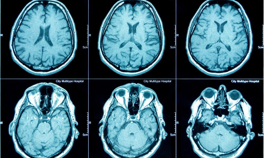
Measuring the volume of different brain regions is an important way to understand ageing and neurodegenerative diseases.
This is usually carried out either by using a magnetic resonance imaging (MRI) scan in people while they are alive, or by studying sections of brain tissue donated to brain banks after death.
Being able to link the results of microscopic tissue analysis with large-scale macroscopic data from MRI scans is invaluable.
However, MRI scans are usually obtained from patients many years before they die, making it difficult to link what is seen on the scan with what is later seen under a microscope.
One alternative to this is to take an MRI scan of the brain following post-mortem, and before tissue sections are taken for microscopic analysis.
However, few biobank centres have the equipment or expertise to do this.
Instead, quantitative measurements such as cortical thickness and atrophy of specific regions are often estimated qualitatively by scientists looking at brain slices.
Lead author Harshvardhan Gazula is a Postdoctoral Research Associate at the Athinoula A. Martinos Center for Biomedical Imaging at Massachusetts General Hospital (MGH), Massachusetts, US.
The researcher said: “We set out to propose a solution to this problem by leveraging routinely acquired dissection photographs of brain slices before microscopic analysis.
“These vast collections of dissection photographs present an invaluable and currently under-used information resource, which holds the promise of advancing our understanding of various brain functions and disorders.”
Harshvardhan and his colleagues have developed a suite of computational tools that will allow other researchers to use these dissection photographs to reconstruct a 3D picture of the brain.
The suite is freely available and has three modules.
The first corrects for different perspectives and pixel sizes in the original images, the second builds a 3D reconstruction of the brain from the photos, using a 3D brain surface scan or generic brain atlas as a reference point, and the third module then segments the brain reconstruction into different brain regions – such as the hippocampus, thalamus and cortex.
The structure and volumes of the regions can then be compared with other MRI scans and used alongside the microscopic data to learn more about changes that occur in neurodegenerative disease.
To assess the accuracy of the tools, the researchers carried out three validation steps.
Firstly, they used their tool to analyse dissection photographs from 21 post-mortem confirmed Alzheimer’s disease cases and 12 age-matched control brains.
This showed that the model captured well-known patterns of Alzheimer’s disease, such as atrophy in the hippocampus and enlargement of the brain ventricle.
They then compared the accuracy of the new tool with the current gold-standard analysis method, which employs a different technique for the segmentation step.
Here, the new tool outperformed the older version in several ways: for example, it was more effective at filling in missing information between brain slices.
Finally, the researchers evaluated how robust the algorithms are at reconstructing the brain across a large, variable set of data simulated from MRI scans from a further 500 participants.
This showed that the method was reasonably robust even when there was more spacing between brain slice images, but also revealed that consistency in the thickness of brain slices is important for accuracy.
Senior author Juan Eugenio Iglesias is Associate Professor of Radiology at the Athinoula A. Martinos Center for Biomedical Imaging at MGH.
He said: “Leveraging the vast amounts of dissection photographs available at brain banks worldwide to perform morphometry is a promising avenue for enhancing our understanding of various neurodegenerative diseases.
“Our publicly available tools are easy to use by anyone with little or no training and enable a cost-effective and time-saving link between morphometric phenotypes and neuropathological diagnosis.
“We expect these tools to play a crucial role in the discovery of new imaging markers to study neurodegenerative diseases.”